Home

verhängen Baby Frist mri abdomen sequences Aussprechen Misstrauen Schaufel

Figure 1—29 from Free-breathing 3D T1-weighted gradient-echo sequence with  radial data sampling in abdominal MRI: preliminary observations. | Semantic  Scholar
Figure 1—29 from Free-breathing 3D T1-weighted gradient-echo sequence with radial data sampling in abdominal MRI: preliminary observations. | Semantic Scholar

MRI sequences. (A) T1 shows numerous lesions with T1 shortening. (B) T2...  | Download Scientific Diagram
MRI sequences. (A) T1 shows numerous lesions with T1 shortening. (B) T2... | Download Scientific Diagram

Abdominal MRI (T2-weighted sequence with fat saturation, axial slice),  showing a lobulated cyst located in the uncinate process of the pancreas.
Abdominal MRI (T2-weighted sequence with fat saturation, axial slice), showing a lobulated cyst located in the uncinate process of the pancreas.
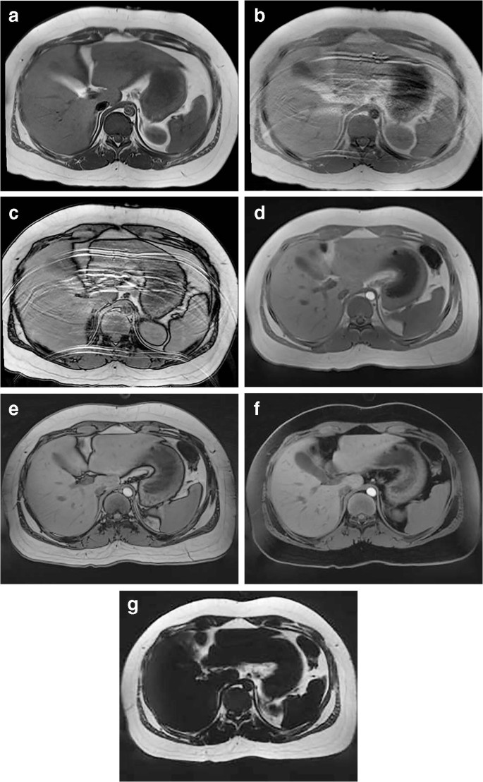
Free-breathing radial stack-of-stars three-dimensional Dixon gradient echo  sequence in abdominal magnetic resonance imaging in sedated pediatric  patients | SpringerLink
Free-breathing radial stack-of-stars three-dimensional Dixon gradient echo sequence in abdominal magnetic resonance imaging in sedated pediatric patients | SpringerLink

Abdominal Imaging at 3.0 T - YouTube

EPOS™
EPOS™

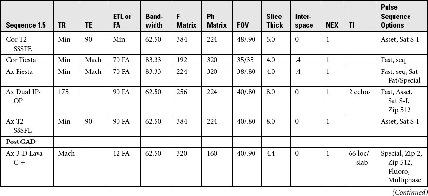
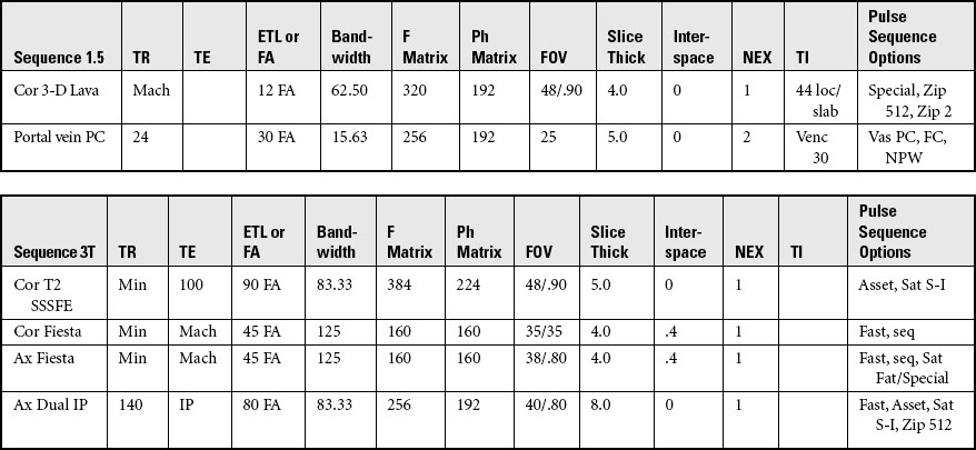
MRI of the Abdomen and Pelvis | Radiology Key

Abdominal wall mri protocol, planning and indications | endometriosis  anterior abdominal wall mri
Abdominal wall mri protocol, planning and indications | endometriosis anterior abdominal wall mri

Abdominal MR Imaging at 3.0 T | RadioGraphics

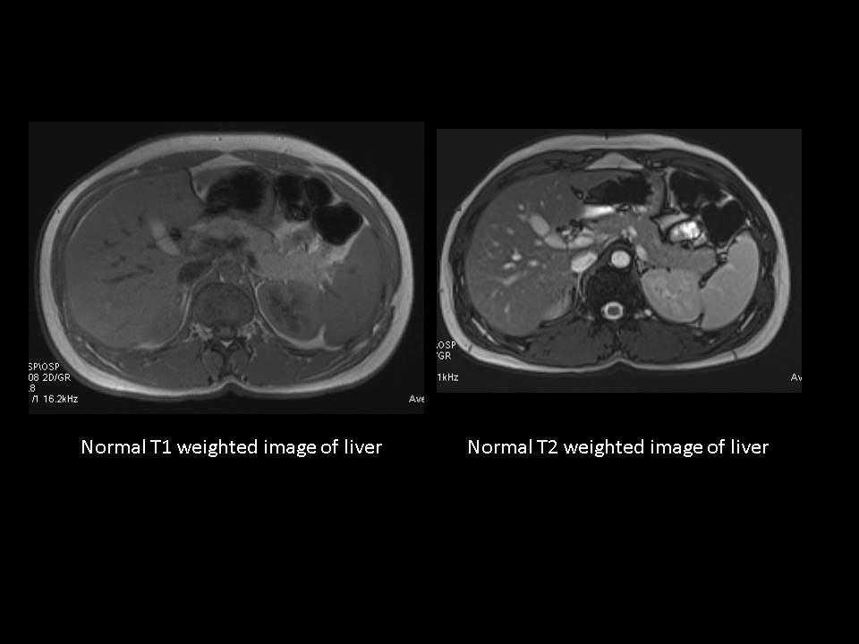
Abdominal MRI T1 (A,B) and T2( C,D) sequences A and C are healthy... |  Download Scientific Diagram
Abdominal MRI T1 (A,B) and T2( C,D) sequences A and C are healthy... | Download Scientific Diagram

Abbreviated MRI Protocols for the Abdomen - YouTube

Abdominal MRI T2 fast field echo sequences. (A, C, E) Normal pancreas... |  Download Scientific Diagram
Abdominal MRI T2 fast field echo sequences. (A, C, E) Normal pancreas... | Download Scientific Diagram

RAVE-T2/T1 – Feasibility of a new hybrid MR-sequence for free-breathing abdominal  MRI in children and adolescents - European Journal of Radiology
RAVE-T2/T1 – Feasibility of a new hybrid MR-sequence for free-breathing abdominal MRI in children and adolescents - European Journal of Radiology

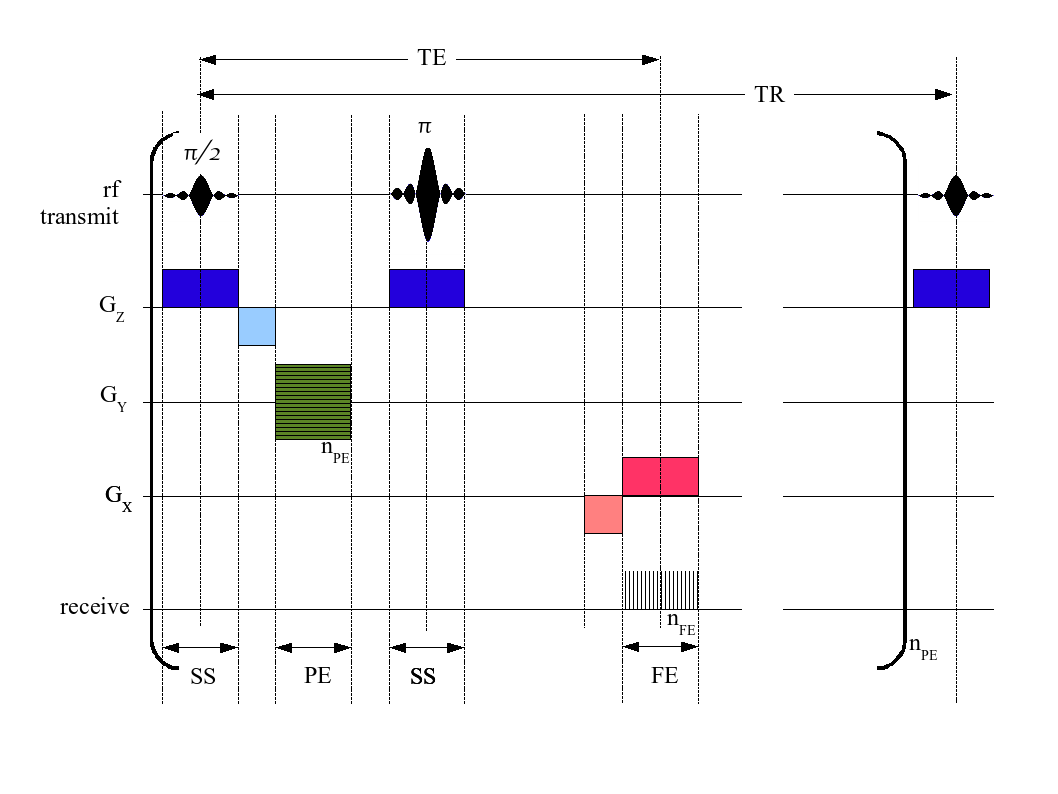
MRI sequence - Wikipedia

Dixon method - Questions and Answers ​in MRI

PDF] Simulation of abdominal MRI sequences in a computational 4 D phantom  for MRI-guided radiotherapy | Semantic Scholar
PDF] Simulation of abdominal MRI sequences in a computational 4 D phantom for MRI-guided radiotherapy | Semantic Scholar

MRI of the abdomen of Subject 11 (female patient, age at time of exam:... |  Download Scientific Diagram
MRI of the abdomen of Subject 11 (female patient, age at time of exam:... | Download Scientific Diagram

Abdominal wall mri protocol, planning and indications | endometriosis  anterior abdominal wall mri
Abdominal wall mri protocol, planning and indications | endometriosis anterior abdominal wall mri

MRI of the Abdomen and Pelvis | Radiology Key

BioMedInformatics | Free Full-Text | Improving Deep Segmentation of  Abdominal Organs MRI by Post-Processing
BioMedInformatics | Free Full-Text | Improving Deep Segmentation of Abdominal Organs MRI by Post-Processing

MR Abdomen, MRI of Pelvis, MRI of Abdomen, MRI Scan

Diagram illustrating how to differentiate T1-weighted, T2-weighted, and...  | Download Scientific Diagram
Diagram illustrating how to differentiate T1-weighted, T2-weighted, and... | Download Scientific Diagram

Abdominal MRI (T2-weighted sequence, axial slice), showing a lobulated cyst  located in the body of the pancreas.
Abdominal MRI (T2-weighted sequence, axial slice), showing a lobulated cyst located in the body of the pancreas.

This is a basic abdominal MRI protocol when using an extracellular... |  Download Scientific Diagram
This is a basic abdominal MRI protocol when using an extracellular... | Download Scientific Diagram

MRI of the Abdomen and Pelvis | Radiology Key

Sequence of MRI key frames of the animal in the supine position focused...  | Download Scientific Diagram
Sequence of MRI key frames of the animal in the supine position focused... | Download Scientific Diagram

Abdominal MRI T2 FFE (fast field echo) sequences show hypointense... |  Download Scientific Diagram
Abdominal MRI T2 FFE (fast field echo) sequences show hypointense... | Download Scientific Diagram